
Retina
The retina is the light-sensitive
tissue on the inside and back of the eye.
Vision originates in the retina,
which contains photoreceptor cells that convert light into electrical impulses.
These impulses are the visual
information or “pictures” that travel to the brain via the optic nerve.
Most retinal disorders involve a
disruption in the transmission of these impulses.
There are many kinds of retinal
disorders, with a wide variety of causes and symptoms.
What are the symptoms of retinal disorders?
Symptoms include:
What are the symptoms of retinal disorders?
Symptoms include:
- a white pupil
- a loss or partial loss of vision
- night blindness
- a shower of black floaters in vision
- sudden, persistent flashing lights
- an intolerance of light
Tests for evaluation of retinal diseases
Apart from the
routine tests that most ophthalmologists and retinal specialists perform, like visual acuity testing, intraocular pressure check and slit lamp examination, here is a list of tests
that an expert may advise for a detailed retinal evaluation.
- Amsler Grid test
- Indirect Ophthalmoscopy
- Colour Vision Test
- Visual Field Test
- Optical Coherence Tomography
- Fundus Fluorescein Angiogram
- Indo-Cyanine Green Angiography (ICG)
- Ultrasound
- Electroretinogram (ERG)
- Electroculogram (EOG)
- Visual Evoked Response
 Common eye conditions that affect the retina
include
Common eye conditions that affect the retina
include
DIABETIC RETINOPATHY
Diabetes affects the eye in various
ways.
- Diabetic retinopathy
- Cataract
- Glaucoma
Cataract requires cataract surgery and implant of an artificial lens (or intraocular lens), while glaucoma refers to increase in pressure of the fluid inside the eye (aqueous) that leads to optic nerve damage and loss of vision. A person with diabetes is nearly twice as likely to get glaucoma as other adults.
 Diabetic
retinopathy refers to damage to the retina caused by abnormal blood flow
related to diabetes mellitus, which can potentially lead to severe loss of
vision.
Diabetic
retinopathy refers to damage to the retina caused by abnormal blood flow
related to diabetes mellitus, which can potentially lead to severe loss of
vision.
In
some people with diabetic retinopathy, blood vessels may swell and leak fluid.
In other people, abnormal new blood vessels grow on the surface of the retina.
The retina is the light-sensitive tissue at the back of the eye. A healthy
retina is necessary for good vision.
Diabetic retinopathy usually affects both eyes.
Remember:
- If you have diabetes get a
comprehensive dilated eye exam at
least once a year.
- Proliferative retinopathy can develop without symptoms. At this advanced stage, you are
at high risk for vision loss.
- Macular
edema can develop without symptoms
at any of the four stages of diabetic retinopathy. You can develop both
proliferative retinopathy and macular edema and still see fine. However,
you are at high risk for vision loss..
- Whether or not you have
symptoms, early detection and
timely treatment can prevent vision loss.
Treatment for Diabetic Retinopathy:
The
usual treatment paradigms for the disease are:
- Laser
treatment
- Anti-VEG-F
treatments
- Retinal
Surgery
 Macular Degeneration
Age-related macular degeneration (AMD) presents
as difficulty in reading small print, or seeing faces, leading to progressive
loss of central vision. AMD is the most common cause of vision loss in
individuals over 55 years of age.

There are two types of macular degeneration;
·
‘wet’ type (about 10% of cases) that can lead to severe loss
of vision and needs treatment in most cases, and
·
‘dry’ type seen in about 90% of cases that does not
usually progress to significant visual loss.
Treatment:
Dry AMD: No medical or surgical treatment is available at this time. However, vitamin supplements with antioxidants, lutein and zeaxanthin, have been suggested by a National Eye Institute, USA sponsored study.
Wet AMD: Anti-angiogenics or anti-VEGF (anti-Vascular Endothelial Growth Factor) agents have been shown to be effective in causing regression of the abnormal blood vessels and improvement of vision when injected directly into the vitreous cavity of the eye.
The injections have to be repeated monthly or in
six weeks.
Agents such as Ranibizumab (Lucentis),
Bevacizumab (Avastin), and Pegaptanib (Macugen) are routinely used these days.
Only Ranibizumab and Pegaptanib are approved by the FDA for AMD, though
Bevacizumab is used off-label.
Photodynamic therapy has also been used to treat
wet AMD.
RETINAL DETACHMENT

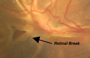 Retinal detachment is a condition when the retina separates away from its normal
position. This can happen at any age, and can lead to sudden or slow loss
of vision, depending on the actual cause.
Retinal detachment is a condition when the retina separates away from its normal
position. This can happen at any age, and can lead to sudden or slow loss
of vision, depending on the actual cause.
The detachment can occur due to many causes,
 including
a hole developing in the retina, as sometimes happens in myopia,
including
a hole developing in the retina, as sometimes happens in myopia,
pull or
traction developing on the retina as can happen in diabetic retinopathy, or
injury
to the retina. In most cases, surgery is
required to correct this disorder, which should be undertaken sooner to
prevent further detachment and potential visual impairment.
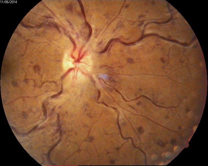
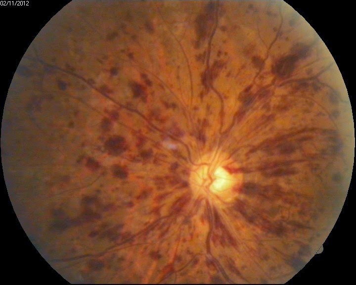
RETINITIS PIGMENTOSA (RP) NIGHT BLINDNESS
Retinitis pigmentosa (RP) refers to a group of inherited diseases that lead to slow and progressive retinal degeneration that causes damage to the
important cells of the retina, the rods and the cones.
People
with RP usually experience night
blindness early on, and later may develop
progressive and gradual reduction in their vision and field of view.
The
diagnosis of retinitis pigmentosa relies upon documentation of progressive loss
in photoreceptor cell function by electroretinography
(ERG) and visual field testing.
The mode of inheritance of RP is determined by family
history. RP can be inherited in an
autosomal dominant, autosomal recessive, or X-linked manner. X-linked RP can be
either recessive, affecting primarily only males, or dominant, affecting both
males and females, although males are usually more mildly affected.
Genetic
counseling depends on an accurate diagnosis, determination of the mode of
inheritance in each family, and results of molecular genetic testing.
Treatment:
·
There is no effective treatment for this condition at this time.
·
Wearing sunglasses to
protect the retina from ultraviolet light may help preserve vision.
·
Some studies have suggested that
treatment with antioxidants (such
as high doses of vitamin A palmitate) may slow the disease in some patients
RETINOPATHY OF PREMATURITY (ROP)
Retinopathy
of Prematurity (ROP) affects the retina in premature babies who are born weighing
less than about 2.75 pounds (1250 grams) or those born before 31 weeks of
pregnancy. This disorder usually affects both eyes at the same time, and is
one of the most common causes of visual impairment in childhood.
CANCERS IN RELATION TO THE RETINA
There are two types of cancer in the eye;
Retinoblastoma, which is more
common among children,
If the child’s eye looks ‘white’ or appears
misaligned, it may mean an abnormality in the eye and this must be
investigated. Unfortunately, retinoblastoma can affect both eyes simultaneously
Malignant Melanoma, which tends
to strike older age groups.
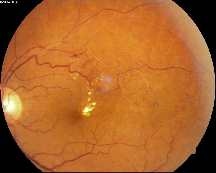
RETINAL VEIN OCCLUSION
Central retinal vein occlusion
(CRVO) is a blockage of the main vein in the retina.
Blockage of the branches of the central vein of the retina is called branch retinal vein occlusion (BRVO).
Blockage of the branches of the central vein of the retina is called branch retinal vein occlusion (BRVO).
 The blockage causes the walls
of the vein to leak blood and excess fluid into the retina which leads to
blurring of vision. When the leakage is in the central part of the retina, the
vision loss is marked.
The blockage causes the walls
of the vein to leak blood and excess fluid into the retina which leads to
blurring of vision. When the leakage is in the central part of the retina, the
vision loss is marked. 
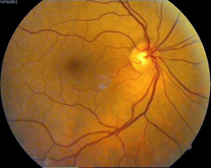
CENTRAL SEROUS RETINOPATHY (CSR)
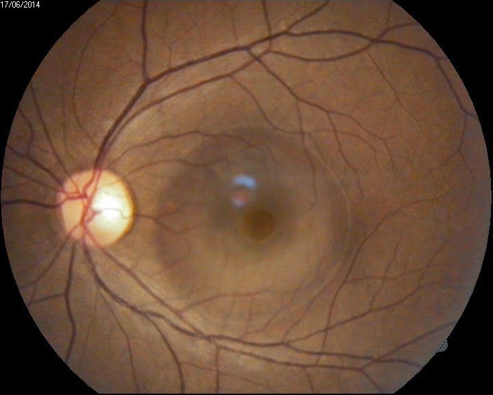 CSR is a condition which causes
temporary or permanent impairment of vision. It usually affects men between 20 – 45 years of age and is associated with
stress.
CSR is a condition which causes
temporary or permanent impairment of vision. It usually affects men between 20 – 45 years of age and is associated with
stress.A small pool of fluid accumulates under the macula (part of the retina). This typically becomes noticeable when central vision is affected.